-
Upconverting Nanoparticles
Oct 24, 2017 | ACS MATERIAL LLCUpconverting nanoparticles have attracted a large amount of interest due to their ability to partake in bioimaging and therapy applications. Upconversions display anti-Stokes shifts in which the summed, single emitted photon has a higher energy than the incident photons. While this is an unnatural phenomenon, rare earth metals have been used to assist in upconversions due to its multiple excitation levels. With this ability, these nanoparticles can be used to possibly overcome limitations (as found in quantum dots) when displaying fluorescent properties. In this article, we will discuss the process of upconverting nanoparticles and how it can potentially provide a better alternative in biological and imaging fields.
Introduction
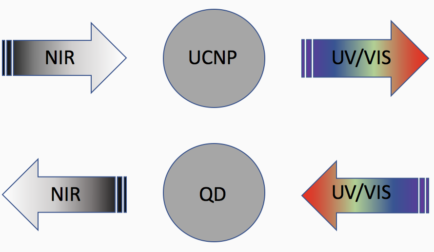
Upconverting nanoparticles (UCNPs) are luminescent nanomaterials that range between 1 to 100 nm in size. Conventional fluorescence typically absorbs a higher energy photon and emits a lower energy photon, but UCNPs does the exact opposite in which it absorbs two or more lower energy photons of a longer wavelength and emits a single higher energy photon of a shorter wavelength. This process can be described by anti-Stokes shifts where the emitted photon has a higher energy than the incident photons. Here, photon emission occurs in the visible or ultraviolet light range of the electromagnetic spectrum and absorption happens in the infrared region.

Figure 1. Emission of a photon (UV or visible light) is higher than that of the incident photon (NIR) for UCNPs whereas QDs emit photons with a lower energy than the incident photons.
Typically, using high energy excitation light has some limitations such as DNA damage, cell death due to long-term irradiation, a large amount of auto-fluorescence from biological tissues resulting in low signal-to-background ratio and short penetration depth for biological tissues. In contrast, upconversion fluorescence involves infrared excitation that is less harmful to cells, reduces auto-fluorescence from biological tissues and penetrates tissues at a greater depth.1 Near infrared (NIR) light is easier to produce in the form of laser radiation when compared to visible and ultraviolet light. This light region is invisible to humans and can reach deep into soft tissues, muscles and bones in the human body, but it also works for applications in displays, photovoltaics, data storage and cancer therapy.
UCNPs Synthesis
NIR light has insufficient photon energies that makes it difficult to apply different techniques to the applications mentioned earlier. Upconversion is an unnatural phenomenon and requires a specific method in order to make this happen. In this case, spectral converters are needed to convert NIR light into higher energy photon emissions.2 Rare earth metals, or lanthanides, have been used in upconversion energy transfer to provide upconversion luminescence and exhibits f-f transitions. Lanthanide-doped nanoparticles can be excited with NIR light, making this cooperation ideal to reduce auto-fluorescence of biological samples and improve image contrasts.
Upconversions tend to work well with lanthanide-doped transition metals ranging from lanthanum to lutetium, as well as scandium and yttrium. The energy levels from the 4f electron configuration from the rare earth metals are copious, which allows for many intraconfigurational transitions. Due to their many energy levels, rare earth ions are known to be promising luminescent centers.3 Upconversions occur with two common processes in lanthanide-doped nanomaterials – excited state absorption (ESA) and energy transfer upconversion (ETU).
The ESA process involves rare earth ions with multiple energy levels that undergoes the absorption of two or more low-energy photons resulting in the transition from ground to excited state and going even further to a much higher excited state. From there, the high-energy photons can be released within these transitions but the absorption cross section of the excited ions needs to be suitable enough to absorb a second pump photon, the necessary additional energy being transferred from another laser-active ion undergoing non-radiative de-excitation.3 This process is commonly used when dopant concentrations are low and energy is least likely to transfer.
For the ETU process, two types of luminescent centers (sensitizer and activator) and a host lattice are used in an energy transfer from singly excited ions (sensitizers) to an emitted ion (activators) and involves an excitation to a higher level from a laser-active ion that would absorb a pump photon. This energy transfer starts with a sensitizer to an excitation from an activator and continues with an even higher excitation to a final fluorescing state. Yb3+ is typically used as a sensitizer due to a large absorption cross-section in the NIR region and it can transfer its energy to activator ions such as Er3+, Tm3+, Pr3+ or Ho3+. The host lattice needs to accommodate the sensitizers and activators but should also have low phonon energies, high chemical stability and low non-radiative energy losses for upconversion emission efficiency.4
Quantum Dots
Lanthanide complexes and UCNPs exhibit unique photo-physical properties and provide a higher signal-to-noise ratio compared to quantum dots.4 Quantum dots (QDs) have been compared to UCNPs as they are also known to have fluorescent properties. They are tiny semiconducting nanocrystals that converts incoming energy into light in pure colors depending on their shape and size. Larger QDs emit longer wavelengths in the red color region while smaller dots emit shorter wavelengths with a blue color. Changing its size can be manipulated and the dots can therefore emit any color of light from the same material.
However, not all QDs are alike, so delivering certain types of QDs inside of cells without destroying them and, in some cases, toxicity of the cells can an issue. The dots have also been shown to reside in cells for weeks to months and can possibly present issues in the body. The two commonly used constituent metals in QD core metalloid complexes, cadmium and selenium, have been known to cause acute and chronic toxicities in vertebrates and considerably in human health. In addition, intermittent emission/blinking can cause the dots to become invisible.5,6 This could mean that the ratio of the emitted energy to the absorbed energy is relatively low and therefore, the low transmittance may remain undetectable or require high-sensitivity detection systems. Nevertheless, QDs are still advantageous phenomenon that have gained high interested due to its tunable properties that are beneficial to various imaging and optical applications.
UCNPs Applications
UCNPs are found to be great optical probes for photoluminescence bioimaging since they are free from many disadvantages attributed to other photoluminscent probes, namely broad emission and susceptibility to bleaching for organic luminophores, blinking and toxicity for semiconductor QDs and so on.7 Since there is a significant advantage with UCNPs, it has been applied to biomedical imaging since the NIR light region reaches deep into cells and bones with little to no damage to cells. These nanoparticles use an anti-Stokes process that utilizes lower energy from NIR light, allowing for a deeply penetrating yet less damaging light source. In fact, toxicity of these UCNPs have been comprehensively reviewed recently, describing their cellular uptake, cytotoxicity, bio-distribution and in vivo excretion while showing much less toxicity in vitro and in animal models than QDs. UCNPs provide a narrow emission spectra, high chemical stability, long luminescence lifetime and high resistance to photoquenching and photobleaching.8
Photodynamic therapy (PDT) delivery systems that can release therapeutic agents to a specific site is another promising application for UCNPs. PDT is an approach that offers drug delivery control by use of an external photon source to allow active therapeutic release to a target area. Since NIR radiation has greater tissue penetration depth compared to UV wavelengths, UCNP drug release has the potential to offer an attractive approach for targeted delivery in vivo.9
There are three different methods to build these drug delivery systems: first, hydrophobic drugs that are encapsulated into “hydrophobic pockets” on the UCNP surface using the hydrophobic-hydrophobic interaction between hydrophobic ligand on the particle surface and the drugs; secondly, drugs are deposited in the pores of mesoporous silica shells coated onto the surface of the UCNPs, accommodating the large amount of drugs; finally, drugs are loaded into a hollow UCNP with a mesoporous shell since it can enable adequate levels of drug loading while maintaining imaging ability. UCNPs can locally trigger UV-activated compounds when irradiated with benign IR irradiation. They can absorb IR light and emit visible light to trigger a photosensitizer that can produce highly reactive singlet oxygen to destroy tumor cells.10
Fluorescence-based optical biosensors are one of the largest group of sensors at the moment and the use of NIR light as an excitation source for biosensing and bioimaging has gained attention due to its optical transparent window in biological tissues. Thus, fluorescence is the source of contrast for most optical bioimaging and the ideal fluorescent probes with strong luminescence, desirable excitation and emission properties and photostability are essential for quality imaging. Since UCNPs have unique advantages, it has been used for multimodal imaging, multifunctional theranostics and optical bioimaging.11
Conclusion
UCNPs have unique optical and fluorescent properties and has currently evolved in a way that can help advance the biological field. This luminescence phenomenon can provide an alternative source to bioimaging, biosensing, in vivo and in vitro applications that has had specific limitations regarding toxicity and blinking. More recent studies have shown these nanoparticles to be effective in obtaining images with a high luminescent lifetime and less damaging to cells. In all, the advancement of UCNPs for more future bio-applications will continue to prove its advantages.
ACS Material Products:
Upconverting Nanoparticles
- UCNPs, Oil Dispersible UCNPs, PEG-Modified UCNPs, Silica-Coated UCNPs, PEG-COOH Modified UCNPs, others.
References
1. Chatterjee, Dev K., et al. “Upconversion Fluorescence imaging of cells and small animals using lanthanide doped nanocrystals.” Biomaterials, Elsevier, 3 Dec. 2007 http://www.sciencedirect.com/science/article/pii/S0142961207008691
2. Chen, Xian, et al. “ChemInform Abstract: Photon Upconversion in Core-Shell Nanoparticles.” ChemInform, vol. 46, no. 21, 2015, doi: 10.1002/chin.201521300.
3. Sun, Ling-Dong, et al. “Upconversion of Rare Earth Nanomaterials.” Annual Review of Physical Chemistry, vol. 66, no. 1, pp. 619-642., doi:10.1146/annurev-physchem-040214-121344.
4. Shashi Bhuckory. Quantum dots and upconverting nanoparticles : Bioconjugation and time- resolved multiplexed FRET spectroscopy for cancer diagnostics. Biotechnology. Universit ́e Paris-Saclay, 2016.
5. Hardman, Ron. “A Toxicologic Review of Quantum Dots: Toxicity Depends on Physicochemical and Environmental Factors.” Environmental Health Perspectives, National Institute of Environmental Health Sciences, Feb. 2006.
6. Nadort, Annemarie, et al. “Quantitative Imaging of Single Upconversion Nanoparticles in Biological Tissue.” PLoS ONE, Public Library of Science, 2013.
7. Chen, Guanying, et al. “Light upconverting core-shell nanostructures: nanophotonic control for emerging applications.” Chemical Society Reviews, The Royal Society of Chemistry, 22 Oct. 2014.
8. Wu, Xiang, et al. “Upconversion Nanoparticles: A Versatile Solution to Multiscale Biological Imaging.” Bioconjugate Chemistry, vol. 26, no. 2, 2015, pp. 166-175., doi: 10.1021/bc5003967.
9. Fedoryshin, Laura L., et al. “Near-Infrared-Triggered Anticancer Drug Release from Upconvering Nanoparticles.” ACS Applied Materials & Interfaces, vol. 6, no. 16, 2014, pp. 13600-13606., doi: 10.1021/am503039f.
10. Chen, Guanying, et al. “Upconversion Nanoparticles: Design, Nanochemistry, and Applications in Theranostics.” Chemical Reviews, vol. 114, no. 10, 2014, pp. 5161-5214., doi: 10.1021/cr400425h.
11. Tan, Guang-Rong, et al. “Small Upconverting Fluorescent Nanoparticles for Biosensing and Bioimaging.” Advanced Optical Materials, 30 May 2016, onlinelibrary.wiley.com/doi/10.1002/adom.201600141/abstract.
