-
BioGraphene & Bio-MoS2 Suspensions in Water
May 15, 2018 | ACS MATERIAL LLCApplications of graphene and its two-dimensional counterparts (MoS2, BN) in the biomedical and healthcare sectors require water dispersions and biocompatible derivatives.1,2 Although the successful exfoliation of these bulk materials using organic solvents,3,4 ionic liquids,5,6 or surfactants7 has been reported, the biocompatibility of the resulting product, quality, expense, and environmental impact still remain unaddressed. 8
Introduction
In an effort to produce biocompatible graphene, proteins were recently used to yield aqueous graphene dispersions using two separate methods, shear and ultrasonication. Sonication gave low exfoliation efficiencies (1 mg mL−1 h−1), limited scalability (100 mL) and induced basal plane defects.9,10 Breaking of the sheets, production of reactive sites and reaction of the damaged sites with oxygen were indicated to be some of the disadvantages of sonication.9 Ultrasonication, for example, was used to exfoliate graphene with bovine serum albumin (BSA).11-13 The protein was suggested to adsorb to graphite platelets and facilitate delamination upon ultrasonication.11-13 The idea was that the adsorbed protein might inhibit restacking of the graphene sheets and contribute to nanoplate stability.13
Alternatively, shear force/turbulence methods were tested in a kitchen blender for the mechanical exfoliation of graphite at rapid exfoliation rates.10,14,15 This method offered low oxidative damage (O > 5%),15 high yields and was mainly used with organic solvents, N-methyl pyrolidine (NMP),10 dimethyl formamide (DMF),15 and some surfactants.14 The shear produced in the turbulent flow exceeded the critical Reynolds number required for delamination (104).15 The results demonstrated that shear generated by a simple kitchen blender is a feasible means for graphene production.14,15
Shear-exfoliation of graphite in a kitchen blender was used along with a variety of edible proteins to produce high concentrations of nearly defect-free graphene aqueous dispersions. By combining the advantages of protein characteristics as exfoliating and stabilizing agents, high quality graphene was produced rapidly. One of the proteins tested for exfoliation efficiency was bovine serum albumin (BSA), an inexpensive edible protein that is a waste product in the meat industry.16 The surface charge of the protein played a critical role in the exfoliation and stabilization of graphene dispersions. This protein-based shear/turbulence exfoliation method proved favorable over sonication or organic solvent methods in terms of yield, quality and water dispersion properties.
BioGraphene & Bio-MoS2
Shear exfoliation of graphite and bulk MoS2 using inexpensive, edible proteins was used for large-scale synthesis of water dispersible, few layer graphene (FLG) and few layer MoS2. These samples were imaged using transmission electron microscopy (Figure 1).
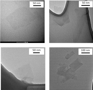
A. TEM Images of BioGraphene
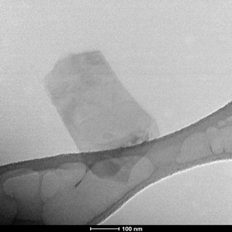
B. TEM Image of Bio-MoS2
Figure 1. A. TEM images of graphene synthesized using bovine serum albumin in a kitchen blender. B. TEM image of a few layered MoS2 sheet synthesized using bovine serum albumin and shear force.
Exfoliation of graphite to high quality graphene with bovine serum albumin (BSA) was achieved using an ordinary kitchen blender. Raman spectroscopy and transmission electron microscopy indicated 3-5 layer, low-defect graphene of 0.5 µm size (Figure 2A). Graphene dispersions dried on ordinary printer paper (650 µg/cm2) showed electrical conductivity of 32,000 S m-1, which is much higher than many graphene/polymer composites (Figure 2B). The BSA-coated graphene was stable under a wide range of pH (3.0-11) and over 5-50 ˚C over two months.
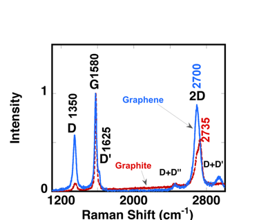
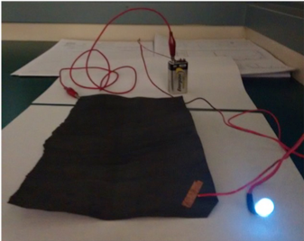
Figure 2. A. Raman spectra of graphene made in a blender (blue) in comparison to graphite (red). B. Electrical conductivity of graphene covered cellulose paper.
MoS2 is a layered material, analogous to graphene, that has exciting applications in electronics, including lithium ion battiers.17,18,19 Shear method with BSA is also effective in exfoliating MoS2 sheets as well as a number of other two-dimensional materials. Our MoS2 shows characteristic Raman peaks at about 384 cm-1 and 409 cm-1 as well as characteristic UV-Visible absorption peaks at 614 nm and 674 nm (Figure 3).20,21
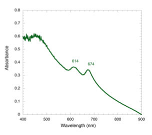
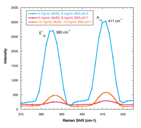
Figure 3. UV-Visible spectrum of a bovine serum albumin exfoliated, water dispersible MoS2 sample, showing characteristic absorption peaks at 614 nm and 674 nm.
Concusions
We have coupled the exciting possibilities of the shear/turbulence method with the surface active properties of edible proteins and produced a multigram scale synthesis of un-oxidized, low-defect graphene and MoS2 suspensions in water. Our suspensions show long-term thermal, pH and storage stability under biologically relevant conditions. These aqueous dispersions with improved biological stability, could be good alternatives to widely used GO or rGO for a variety of applications in biomedical and bioengineering fields.22, 23 Biographene-coated cellulose paper, for example, is unique in that it is a conductive, metal-free cellulose-protein-graphene hybrid material that has not been reported before. The simple exfoliation method reported here could lead to greater access of graphene for laboratories with limited resources or applications that require large quantities, and its use could further revolutionize the graphene industry.
ACS Material Products:
Graphene Series:
Graphene-like Materials:
Quantum Dots & Upconverting Nanoparticles:
References
1. C. Chung, Y. K. Kim, D. Shin, S. R. Ryoo, B. H. Hong, D.-H. Min, Acc. Chem. Res. 2013, 46, 2211.
2. C. H. A. Wong, Z. Sofer, M. Kubešová, J. Kucˇera, S. Mateˇjková, M. Pumera, Pro. Natl. Acad. Sci. USA 2014, 111, 13774.
3. M. Yi, Z. Shen, J. Mater. Chem. A 2015, 3, 11700.
4. X. Chen, J. F. Dobson, C. L. Raston, Chem. Commun. 2012, 48, 3703. [5] T. Morishita, H. Okamoto, Y.
5. Katagiri, M. Matsushita, K. Fukumori,Chem. Commun. 2015, 15, 12068.
6. I. W. P. Chen, Y. S. Chen, N. J. Kao, C. W. Wu, Y. W. Zhang, H. T. Li, Carbon 2015, 90, 16.
7. M. Lotya, P. J. King, U. Khan, S. De, J. N. Coleman, ACS Nano 2010, 4, 3155.
8. K. Yang, Y. Li, X. Tan, R. Peng, Z. Liu, Small 2013, 9, 1492.
9. T. Skaltsas, X. Ke, C. Bittencourt, N. Tagmatarchis, J. Phys. Chem. C 2013, 117, 23272.
10. K. R. Paton, E. Varrla, C. Backes, R. J. Smith, U. Khan, A. O’Neill, C. Boland, M. Lotya, O. M. Istrate, P. King, T. Higgins, S. Barwich, P. May, P. Puczkarski, I. Ahmed, M. Moebius, H. Pettersson, E. Long, J. Coelho, S. E. O’Brien, E. K. McGuire, B. M. Sanchez, G. S. Duesberg, N. McEvoy, T. J. Pennycook, C. Downing, A. Crossley, V. Nicolosi, J. N. Coleman, Nat. Mater. 2014, 13, 624.
11. S. Ahadian, M. Estili, V. J. Surya, J. Ramon-Azcon, X. Liang, H. Shiku, M. Ramalingam, T. Matsue, Y. Sakka, H. Bae, K. Nakajima, Y. Kawazoe, A. Khademhosseini, Nanoscale 2015, 7, 6436.
12. D. Joseph, N. Tyagi, A. Ghimire, K. E. Geckeler, R. Soc. Chem. Adv. 2014, 4, 4085.
13. A. M. Gravagnuolo, E. Morales-Narváez, S. Longobardi, E. T. da Silva, P. Giardina, A. Merkoçi, Adv. Funct. Mater. 2015, 25, 2771.
14. E. Varrla, K. R. Paton, C. Backes, A. Harvey, R. J. Smith, J. McCauley, J. N. Coleman, Nanoscale 2014, 6, 11810.
15. M. Yi, Z. Shen, Carbon 2014, 78, 622.
16. P. H. Yun-Hwa, A. O. Jack, in Handbook of Analysis of Edible Animal By-Products, CRC Press, Boca Raton, Florida, USA 2011, pp. 13–35.
17. B. Radisavljevic, A. Radenovic, J. Brivio, V. Giacometti and A. Kis, Nat. Nanotechnol., 2011, 6, 147–150.
18. J. W. Seo, Y. W. Jun, S. W. Park, H. Nah, T. Moon, B. Park, J. G. Kim, Y. J. Kim and J. Cheon, Angew. Chem., Int. Ed., 2007, 46, 8828–8831.
19. G. D. Du, Z. P. Guo, S. Q. Wang, R. Zeng, Z. X. Chen and H. K. Liu, Chem. Commun., 2010, 46, 1106–1108.
20. T. Sekine, C. Julien, I. Samaras, M. Jouanne, M. Balkanski, Mater. Sci. Eng. B 3 (1989) 153.
21. Benavente E, et al. Intercalation chemistry of molybdenum disulfide. Coordination Chemistry Reviews. 2002; 224(1):87–109.
22. Q. Tang, Z. Zhou, Z. Chen, Nanoscale 2013, 5, 4541.
23. A. C. Ferrari, F. Bonaccorso, V. Fal’ko, K. S. Novoselov, S. Roche, P. Boggild, S. Borini, F. H. L. Koppens, V. Palermo, N. Pugno, J. A. Garrido, R. Sordan, A. Bianco, L. Ballerini, M. Prato, E. Lidorikis, J. Kivioja, C. Marinelli, T. Ryhanen, A. Morpurgo, J. N. Coleman, V. Nicolosi, L. Colombo, A. Fert, M. Garcia-Hernandez, A. Bachtold, G. F. Schneider, F. Guinea, C. Dekker, M. Barbone, Z. Sun, C. Galiotis, A. N. Grigorenko, G. Konstantatos, A. Kis, M. Katsnelson, L. Vandersypen, A. Loiseau, V. Morandi, D. Neumaier, E. Treossi, V. Pellegrini, M. Polini, A. Tredicucci, G. M. Williams, B. Hee Hong, J.-H. Ahn, J. Min Kim, H. Zirath, B. J. van Wees, H. van der Zant, L. Occhipinti, A. Di Matteo, I. A. Kinloch, T. Seyller, E. Quesnel, X. Feng, K. Teo, N. Rupesinghe, P. Hakonen, S. R. T. Neil, Q. Tannock, T. Lofwander, J. Kinaret, Nanoscale 2015, 7, 4598.